Urology Surgery Research
Head
|
|
|
Prof. Daniel Eberli, MDDirector Department
|
|

Organization
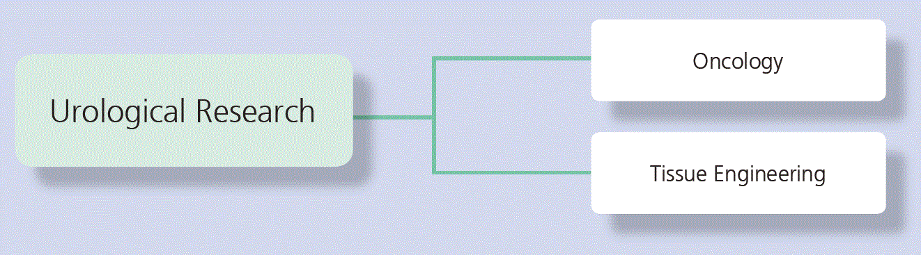
The department of Urology focuses its research interests on the two areas Uro-Oncology and Tissue Engineering/Regenerative Medicine.
Focus Prostate Cancer Studies
Based on our previously submitted patent on “Novel Urine Biomarker for Prostate Cancer”, we perform a pre-validation study testing the clinical relevance of the biomarker for diagnosis and prognosis of prostate cancer (PCa) and the possibility to substantially reduce the number of unnecessary biopsies.
As an alternative biomarker type we explore for PCa diagnostics, extracellular vesicles (EVs) are particularly intriguing. EVs, which are membrane-bound nanoparticles secreted by cells into the blood or media, are known to carry essential specific to their cell of origin. Using advanced in vitro models, we harvested EVs produced by PCa cells and analyzed them with proteomics (LC-MS/MS) to determine which cargo are related to disease progression. Candidate proteins are being validated in retrospective analysis of patient samples from the proCOC (prostate cancer outcome study) biobank.
Multiple androgen receptor (AR) dependent and independent resistance mechanisms limit the efficacy of current treatment modalities for castration resistant prostate cancer (CRPC). Autophagy is a survival mechanism in cells exposed to anti-cancer treatment. We hypothesized that also a promising N-terminal or C-terminal targeting-AR treatment may lead to up-regulation of autophagy, which can be targeted by a combination therapy with autophagy inhibitors. Current research focuses on in vitro and in vivo studies investigating the antitumor effect of a double-treatment using autophagy and AR inhibitors.
PCa is responsible for the second most cancer-related deaths in men after lung cancer in Switzerland. Precise visualization and therapy of primary and recurrent PCa foci is one of the prominent challenges in these tumor patients. Prostate-specific membrane antigen (PSMA) based imaging and therapy is increasingly used for targeted PCa management. However, a low PSMA surface expression in patients with low-volume and low-grade cancer can limit accurate imaging and therapy. In vitro and in vivo data has demonstrated that androgen deprivation therapy (ADT) induces PSMA surface expression. However, ADT might negatively influence disease progression in certain patients. We hypothesize that upregulation of PSMA expression can also be induced by other commonly used FDA-approved compounds indirectly targeting the AR pathway. We aim to identify these pharmacological compounds inducing the PSMA expression in vitro and in vivo.
We participate in several academic international randomized controlled trials and prospective studies (REDUSE, IMPROVE, PEACE III, PBCG) to improve outcomes in patients with advanced PCa. As a first results of the international multicenter PBCG (prospective biopsy collaboration group) study a new risk calculator for PCa and has been published 2018 (“A Contemporary Prostate Biopsy Risk Calculator Based on Multiple Heterogeneous Cohorts”, Eur Urol 2018). We are currently gathering funding for a phase I and II trial on bipolar androgen therapy (BAT) with a new medical compound in advanced PCa patients. The aim is to identify which PCa subtypes may respond to BAT and find predictive markers. For future biomarker discovery, we established the two biobanks proCOC and metaPROC (metastatic prostate cancer).
Focus Testicular Cancer Studies
As participating institution in the Swiss Austrian German Testicular Cancer Cohort Study (SAG TCCS) we are studying the quality of life and follow-up schedules in patients with testicular cancer. In addition, we are currently conducting a research project funded by the Swiss Cancer League to define the role of microRNAs in the management of testicular cancer patient surveillance. Additionally our group contributes clinical patient data to several international collaborations to optimize clinical care.
Urologic Tissue Engineering
Targeting urologic diseases such as urinary incontinence, the Tissue Engineering group is following different approaches to grow stem cells and initiate tissue regeneration.
In a first approach, we use human skeletal muscle precursor cells (MPCs) for tissue (re)generation. We coordinate an international consortium of the Horizon 2020 EU program and a project entitled Multisystem Cell Therapy for Improvement of Urinary Incontinence (MUSIC) (www.music2020.ch). In this first phase clinical trial, we will treat 40 patients with their autologous muscle precursor cells. Patient-specific cell batches are produced in clean room facilities under GMP conditions. In combination with post injection electromagnetic stimulation, we expect an improved regeneration of the sphincter muscle.
A second approach uses adipose derived stem cells (ADSC) as a key instrument to bioengineer contractile bladder tissue. The ADSCs can differentiate to smooth muscle cells (SMC) under natural conditions. Their long-term cell fate in vivo however is uncertain. We evaluate different combinations of cells to improve the bladder tissue formation by modification of the microenvironment and the enhancement of cell-to-cell interactions. As autologous SMC cannot be harvested from organs with end-stage disease and tissue regeneration requires large amount of functional SMC, there is an urgent need for other cell sources. We investigate the functional role of autophagy during differentiation and remodeling of ADSCs to SMC in vitro.
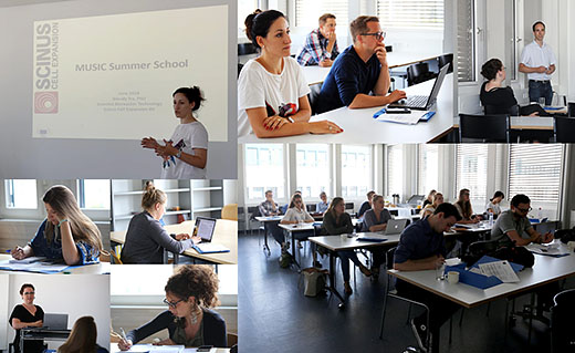
Fig 1: Impressions from summer school 2018
Furthermore, we aim to develop a functional substitute for the improvement of the bladder wall function for patients suffering from end-stage bladder disease. We are investigating the regenerative capabilities of primary bladder SMCs and pre-differentiated, smooth muscle-like ADSCs in compressed collagen hydrogel scaffolds.
A central concern associated with the use of any cell source for tissue engineering is the non-invasive monitoring of in vivo tissue formation. We therefore apply MRI to directly assess stem cell differentiation and skeletal muscle fiber formation. Since the regenerated tissue quality after stem cell therapy is crucially important for its proper function, we apply neuromuscular electromagnetic stimulation (NMES) to support in vivo tissue development, cell survival and innervation.
Collaborations
-
PD Dr. med S. Santourlidis, Heinrich-Heine University, Düsseldorf, Germany
-
Prof. Dr. Michael Detmar, Institute of Pharmaceutical Sciences, ETHZ
-
Prof. Dr. Peter Wild, Senckenberg Institut für Pathologie, Universitätsmedizin Frankfurt, Germany
-
Proteomedix AG, Schlieren
-
University of Applied Science North Western Switzerland (FHNW)
-
Prof. Dr. Arnold von Eckardstein, Institute of Clinical Chemistry
-
Dr. Andrew Vickers, Memorial Sloan Kettering Cancer Center, New York, USA
-
Prof. Dr. Donna Ankerst, Technical University, Munich
-
Prof. Rita Gobet & PD Dr. Maya Horst Division of Pediatric Urology, University Children’s Hospital Zurich
-
Prof. Hans Uwe Simon, Pharmacology Institute; Bern
Prof. Dr. Simon M. Ametamey, Dpt. Pharmaceutical Sciences, ETHZ, Zurich, Switzerland -
Prof. Dr. Christoph Handschin, Biozentrum Basel, Basel, Switzerland
-
PD Dr. med. Andreas Boss, Institute for Diagnostic and Interventional Radiology, USZ
-
Dr. sc. nat. Martin Ehrbar, Division of Obstetrics, University Hospital Zurich